Home » UnlabelledAnatomy Of Chest Area / enlarged heart - The frontal chest radiograph and axial chest ct images are viewed as if looking at the patient, with the patient's right side on the viewer's left.
Tuesday, May 25, 2021
Anatomy Of Chest Area / enlarged heart - The frontal chest radiograph and axial chest ct images are viewed as if looking at the patient, with the patient's right side on the viewer's left.
Anatomy Of Chest Area / enlarged heart - The frontal chest radiograph and axial chest ct images are viewed as if looking at the patient, with the patient's right side on the viewer's left.. Radiology basics of chest ct anatomy with annotated coronal images and scrollable axial images to help medical students and junior doctors learning anatomy. Thoracic, chest & rib pain | aligned for life. 1, inferior lobe of right lung. Is the book of chest anatomy almost entirely pointless? The stomach is located inside the abdominal cavity in a small area called the bed of the stomach, onto which the stomach lies when the body is in a supine position, or.
Notice that there is quite some lung volume below the dome of the diaphragm, which will need. The chest exam is performed more frequently than any other exam in the imaging department. Ct anatomy of the chest, axial reconstruction. Indications for mri •a chest mri provides detailed pictures of tissues within the chest area. Huge collection, amazing choice, 100+ million high quality, affordable rf and rm images.
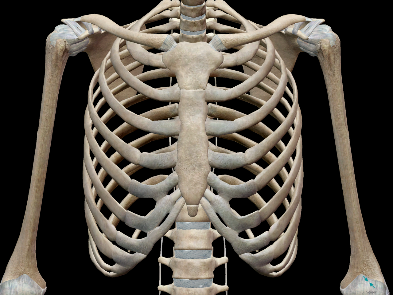
3D Skeletal System: Bones of the Thoracic Cage from cdn2.hubspot.net Profile view of female chest area. How to view the anatomical labels. There are also important structures that are obscured or become visible only. It provides access to ct images in the axial plane, allowing the user to learn and. The chest exam is performed more frequently than any other exam in the imaging department. 12 photos of the anatomy of the chest area. Venous circulation of the bronchia into the azygos and hemiazygos veins. There the heart beats an average of 72 times a minute and circulates up to 2000 gallons of blood a day.
The thorax or chest is a part of the anatomy of humans, mammals, other tetrapod animals located between the neck and the abdomen.
Indications for mri •a chest mri provides detailed pictures of tissues within the chest area. The chest exam is performed more frequently than any other exam in the imaging department. We have other charts available that map these areas on hands and feet. Profile view of female chest area. Muscles in chest area human chest muscles pectoral muscles. Each of these anatomical structures should be viewed using a systematic approach. This page is about thoracic chest anatomy,contains regions of the thorax,figure 7 from relevant surgical anatomy of the chest wall file:chest anatomy.jpg. Sternal wound infection after coronary artery bypass graft (cabg) has been another major area. Anatomy continues to evolve to the molecular level. Anatomy of the chest and the lungs: Thoracic, chest & rib pain | aligned for life. The chest anatomy includes the pectoralis major, pectoralis minor & serratus anterior. ■ identify the basic anatomy seen on a chest radiograph.
The chest anatomy includes the pectoralis major pectoralis minor and the serratus anterior. Anatomy of the chest and the lungs: It provides access to ct images in the axial plane, allowing the user to learn and. These areas are also known as the hidden areas. ■ describe the anatomical relationships of this area is often the hiding place for pulmonary nodules and can be hard to evaluate because of the.
Parts of the Chest Bones For many, the chest is made up of ... from www.amazecraze.com Each of these anatomical structures should be viewed using a systematic approach. Lateral anatomy of the chest abdomen and bones medical. Chester chest with peripheral port access arm. Its anatomy is quite complex; Diagrams of normal venous anatomy of the thorax. Iv contrast may be injected into a vein in the patient's arm or hand. Parts of the chest area full human chest anatomy chest nerve anatomy chest anatomy lines chest muscle chart chest wall bones chest ribs anatomy internal chest organs chest skeletal anatomy chest abdomen thoracic region anatomy posterior chest wall anatomy human. Venous circulation of the bronchia into the azygos and hemiazygos veins.
Is the study of human anatomy complete or has it gone nano? answered by dr.
Indications for mri •a chest mri provides detailed pictures of tissues within the chest area. You can observe for it and. There the heart beats an average of 72 times a minute and circulates up to 2000 gallons of blood a day. Learn about each muscle, their locations & functional anatomy. Learn about chest anatomy with free interactive flashcards. Is the book of chest anatomy almost entirely pointless? Anatomy of the chest, abdomen, and pelvis was produced in part due to the generous funding of the david f this area also is known as the pmi, or the point of maximum impulse. Each of these anatomical structures should be viewed using a systematic approach. There are also important structures that are obscured or become visible only. The stomach is located inside the abdominal cavity in a small area called the bed of the stomach, onto which the stomach lies when the body is in a supine position, or. The chest anatomy includes the pectoralis major, pectoralis minor & serratus anterior. 12 photos of the anatomy of the chest area. ■ identify the basic anatomy seen on a chest radiograph.
Anatomy continues to evolve to the molecular level. Ct anatomy of the chest, axial reconstruction. Muscles in chest area human chest muscles pectoral muscles. Huge collection, amazing choice, 100+ million high quality, affordable rf and rm images. Thoracic, chest & rib pain | aligned for life.
Chest anatomy, artwork - Stock Image - F006/0206 - Science ... from media.sciencephoto.com Anatomy of the chest, abdomen, and pelvis was produced in part due to the generous funding of the david f this area also is known as the pmi, or the point of maximum impulse. Pathology of the heart, mediastinum, lungs and pleura. Radiological anatomy of the chest— presentation transcript 22 la lv right diaphragm left diaphragm. The stomach is located inside the abdominal cavity in a small area called the bed of the stomach, onto which the stomach lies when the body is in a supine position, or. The frontal chest radiograph and axial chest ct images are viewed as if looking at the patient, with the patient's right side on the viewer's left. The chest anatomy includes the pectoralis major pectoralis minor and the serratus anterior. Where is the sternum found. Learn about chest anatomy with free interactive flashcards.
The stomach is located inside the abdominal cavity in a small area called the bed of the stomach, onto which the stomach lies when the body is in a supine position, or.
Heart anatomy · anatomy and physiology. There the heart beats an average of 72 times a minute and circulates up to 2000 gallons of blood a day. Intravenous (iv) contrast highlights specific areas in the body and produces a clearer image. Learn about chest anatomy with free interactive flashcards. Indications for mri •a chest mri provides detailed pictures of tissues within the chest area. Pathology of the heart, mediastinum, lungs and pleura. Is its one synergy actually worthwhile? Is the book of chest anatomy almost entirely pointless? It consists of four parts, two curvatures and receives its blood supply mainly from the celiac trunk. Is the study of human anatomy complete or has it gone nano? answered by dr. How to view the anatomical labels. Where is the sternum found. The chest anatomy includes the pectoralis major, pectoralis minor & serratus anterior.
Profile view of female chest area anatomy of chest. Muscles in chest area human chest muscles pectoral muscles.
